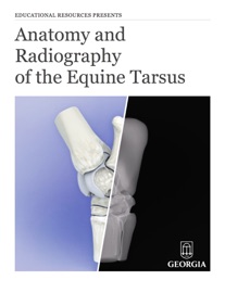
When veterinary students learn anatomy and radiography, they frequently encounter difficulties with the equine tarsus. This is true for a variety of reasons, not the least of which is the number of bones and joints comprising the tarsus, and the apparent overlap of the bones on radiographs. Consequently, many students have difficulty recognizing the different radiographic views used to examine the tarsus. This interactive book combines photographs, radiographs and 3-D models of the bones that comprise the horse’s tarsus in an effort to facilitate understanding of this important joint.